Accelerate to discover

Back to filter

Related topics

Discover the Celloger live-cell analyzers : miniaturized and powerful tools for every Biologist

Curiosis

Apr 5, 2024

Celloger live-cell analyzers, image and analyze your cells in real-time, inside your incubator. With its exceptional...

The chicken chorioallantoic membrane as a low-cost, high-throughput model for cancer imaging

Precision X-Ray

Apr 4, 2024

Here, we assessed the chicken chorioallantoic membrane (CAM) as an alternative to mice for preclinical cancer imaging...

A microthrombus-driven fixed-point cleaved nanosystem for preventing post-thrombolysis recurrence

IVIM Technology

Apr 3, 2024

A thrombin-responsive and fixed-point cleaved Fu@pep-CLipo was developed for highly efficient and precise thrombolysis...

Cytek SpectroFlo Software Version 3.3 has been released!

Cytek Biosciences

Mar 25, 2024

SpectroFlo Software Version 3.3 has been released, enabling new assays with SpectroFlo Software version 3.3, including...

Apr 19, 2024

A 19-color single-tube Full Spectrum Flow Cytometry for the detection of Acute Myeloid Leukemia

Cytek Biosciences

Mar 13, 2024

This recent publication in Cytometry Part A describes the development and comprehensive workflow of a single-tube,...

18F-labeled somatostatin analogs for somatostatin receptors (SSTRs) targeted PET imaging of NETs

MOLECUBES

Mar 11, 2024

A novel 18F-radiolabeled somatostatin analogue, [Al18F]NODA-MPAA-HTA, was synthesized and evaluated for positron...

Discover Yokogawa CellVoyager CQ1: Benchtop High-Content Analysis System

Yokogawa

Mar 8, 2024

Unlike flow cytometers, the CellVoyager CQ1 confocal quantitative image cytometer does not require pretreatment such as...

Apr 19, 2024

Visualization of Tumor-Related Blood Vessels in Small Animals by Photoacoustic 3D Imaging System

Luxonus

Feb 21, 2024

In Vivo Label-Free Observation of Tumor-Related Blood Vessels in Small Animals Using a Newly Designed Photoacoustic 3D...

Show all topics (10)

Label-free, real-time live-cell assays for spheroids

Aug 3, 2017

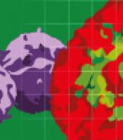
Tumor spheroids exhibit more relevant morphology and increased cell survival compared to 2D cultures and have a hypoxic core. Current methods for assessing the growth and shrinkage of tumor spheroids are limited by one or more of the following: (1) assay workflows are time-consuming, expensive or laborious, (2) a requirement to label the cells (e.g. a fluorescent probe) which may perturb the biology and may not be amenable to primary tissue, (3) single time-point readouts that do not report the full timecourse, (4) indirect readouts (e.g. ATP) that may overlook valuable morphological insight and/or mis-report cell growth.
In this application note we describe methods and validation data for miniaturized (96/384-well) live-cell tumor spheroid assays that are based on non-invasive bright-field image analysis performed with the IncuCyte S3 Spheroid software module. Tumor spheroids are formed in ultra-low attachment (ULA) plates and monitored for size and morphology for up to 2 weeks. These assays are flexible, simple to run and provide automated and direct measures of tumor size in real-time.

Read more
 

Scientific paper
Product news

Get more info

Riccardo Pasculli

Head of application support

Riccardo

Pasculli

+420 731 127 717

Send Message

Brand profile

Sartorius & Essen BioScience

Essen BioScience & Sartorius is a team of engineers and biologists with deep expertise in cell-based assays and biomolecular interaction analysis. They invent, manufacture, supply and support instrumentation, reagents and protocols.

Related products

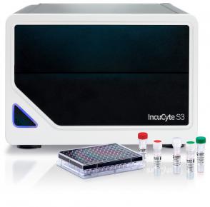
High throughput, real-time and automated measurements of live-cell activity, inside your incubator

show detail

We supply and support Life-Science Technologies in the territory of Central and Eastern Europe.

Czech Republic

Slovakia

Hungary

Poland

Croatia

Slovenia

Serbia

Romania

Bulgaria

Latvia

Lithuania

Estonia

Russia

Ukraine

Belarus

Turkey