Products

“Lago X is an exceptional tool for whole-body applications, with its exceptional reproducibility, signal stability and camera sensitivity, shortening imaging times to less then 1s. Based on our experience, we are able to detect metastases in cancer model much sooner and in more sites than other in-vivo systems. Lago X software is freely available for image processing and quantification, which allows involvement of broad research community into raw data analysis. Finally, we appreciate large field of view, which allows 10 adult mice to be imaged at once. Such features makes Lago X a breakthrough solution for high throughput and high demand in-vivo applications.“
Institute of Molecular Genetics of the Czech Academy of Sciences, Prague, Czech Republic

“The Ami HTX optical imaging system is a flexible tool that we utilize for a range of in vivo and in vitro experiments. The software is intuitive and simple to use. This technology provides a great sensitivity, which is required for our experiments. The system is extremely stable, as there is no external refrigeration system to fail. The hardware is easy to operate and the compact size of the machine saves the working space.”
Medical Faculty, Ostrava,Czech Republic

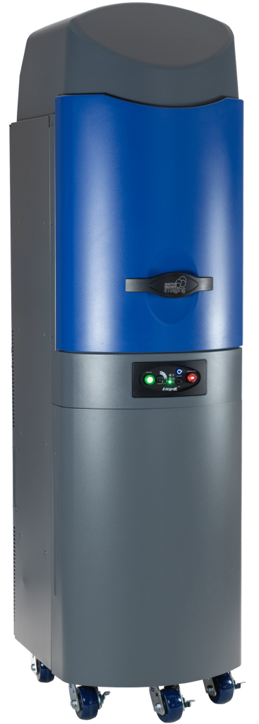
Lago / Lago X – Spectral Instruments Imaging
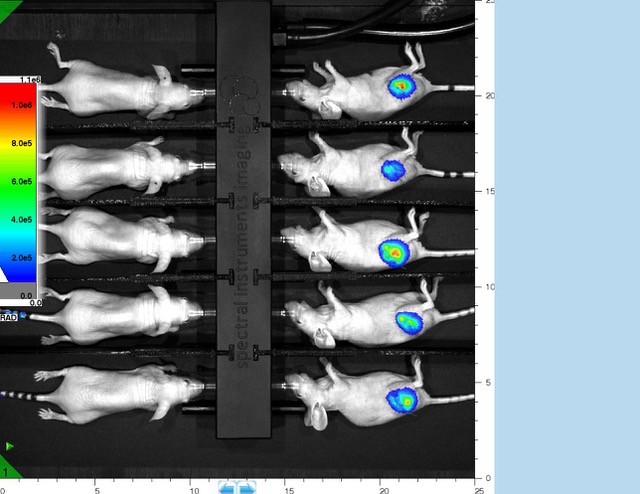
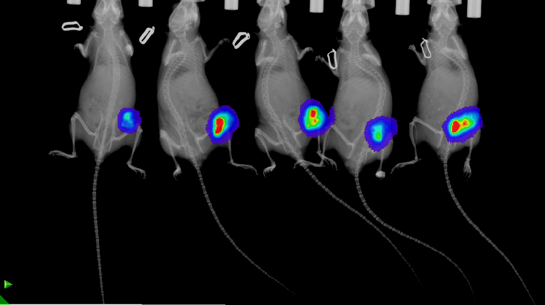
Unprecedented 10 mouse capacity across BLI, FLI and X-Ray
Using Lago and Lago X, researchers in pharma, academia, biotech as well as global CROs can conduct optical imaging system for studying small animal disease model progression, response to therapy, and cell migration in‐vivo.
The cutting edge patented LED based illumination and Faint signal detection provide unprecedented power and previously unattained sensitivity for FLI and BLI. The Lago and Lago X are also suited where early detection and marking disease progression is of value. They come equipped for the largest native field of view (FOV) on the market based on comparable products from other vendors. In fact the Lago and Lago X provides an industry leading 25 cm x 25 cm field of view (FOV) for BLI and FLI. In addition the Lago X provides a 25cm x 22cm FOV for X‐Ray.
This enables the Lago and Lago X to provide an unprecedented and unmatched 10 mouse capacity across BLI, FLI and X-Ray.
The Lago and Lago X are able to deliver unprecedented high throughput capability for critical vaccine research, oncology and other translational studies that also require large sample sizes. The Lago is field upgradeable for X-Ray to the same spec as the Lago X.
The Lago comes equipped with a solid state air‐cooled camera and state of the high performance imaging capabilities. Free, unlimited copies of AMIView software, coupled with competitive support plans, and low entry cost make the Lago the sensible choice for many researchers. The Lago and Lago X are absolutely calibrated.
Key Features:
- Fluorescent imaging
- Bioluminescent imaging
- X‐Ray imaging
- Ultra-high throughput
- 10 mouse capacity for BLI, FLI and X‐Ray
- Pure LED illumination (patented)
- 100X light intensity on specimen
- 14 LED wavelengths from 360 nm to 805 nm
- 20 emission filters included from 490 nm to 870 nm
- Custom emission filters for plant imaging
- Advanced Faint image acquisition
- Solid state cooled CCD camera (–90C), no leaks
- High performance imager
- CCD camera with back illumination
- Ultra‐wide industry leading 25cm x 25cm FOV
- Up to 5 steps from min to max FOV
- X‐Ray FOV is 25cm x 22cm
- 50kV X‐Ray source
- Field upgrade X‐Ray
- Field Upgrade for access port
- Absolutely calibrated
- Acquisition PC and monitor included
- AMIView image analysis software – unlimited free copies
- Vertical sliding door with reduced waste anesthesia gas
- Rigid ‘Strong Back’ high integrity structure
More info at: : https://spectralinvivo.com/imaging-systems/
Discover more
Preclinical Evaluation of 223RaCL2 and Immune Checkpoint Inhibitors in Prostate Cancer Bone MetsReanalyze your in-vivo optical images with Aura SoftwareOptimizing Substrate Dosing for ReliableBioluminescence Imaging (BLI)Come meet us at FELASAOptimizing Substrate Dosing for Reliable Bioluminescence Imaging (BLI)Bruker Lunch Symposium at EMIM 2025 on March 12thDevelopment of Image-Based In Vitro/In Vivo Platform for Targeting CAFsWatch Aura 4.0 Instructional Tutorials – How To Acquire Data Using Easy ModeAutomatic, Real Time Acquisition of Bioluminescent Kinetic CurvesAutomatically Acquire Bioluminescent Kinetic Curve Data in Real Time
Setup a demonstration
with our specialist
Related products


BioFocus newsletter
Register to receive Brand new technology development, Important product updates, Interesting scientific events and more!
Technologies