Accelerate to discover

Back to filter

Related topics

InAlyzer to evaluate G6PD activity in relation with frailty

MEDIKORS

Mar 28, 2023

InAlyzer is body densitometry instrument for lab animals, equipped with 2 X-Ray sources and able to provide valuable...

ACCELA educational webinar: Unlocking Spatial Biology with RareCyte Orion

RareCyte

Mar 16, 2023

Experience Spatial Biology with Orion Multiplex Imaging. Orion is a benchtop, high...

Introducing Cytek Human Leukocyte Kit, the first ever 15-color lyse no-wash assay

Cytek Biosciences

Mar 13, 2023

This kit has been designed to enumerate all major leukocyte subsets and it mirrors and expands on those identified in a...

NanoCellect Webinar: Optimizing the Cell Line Development Process with Microfluidic Cell Sorting

NanoCellect

Mar 3, 2023

Single-cell selection and cloning are required for bioengineering workflows such as antibody production, cell therapy,...

Apr 2, 2023

Flash news - did you know, that you can use Singulator S100 also for plant research?

S2 Genomics

Feb 10, 2023

In this short TechNote you can find how Singulator can help the data quality in Single cell sequencing - now even in...

Advance your Spatial Biology, customize your panel design with RareCyte Orion reagents

RareCyte

Jan 23, 2023

Orion system, the highly multiplexed fluorescent imaging system, in combination with dedicated reagents, empower...

Extensive assessment of Cytokine production on the NovoCyte Advanteon flow cytometer

Agilent technologies

Jan 13, 2023

Cytokines are small molecules essential for immune cell response to activation by pathogens, autoimmunity, or...

Apr 2, 2023

Microfluidic cell sorter sample preparation for genomic assays

NanoCellect

Dec 16, 2022

Single-cell RNA-Sequencing has led to many novel discoveries such as the detection of rare cell populations, microbial...

Show all topics (10)

In-vivo longitudinal study of rodent skeletal muscle atrophy using ultrasonography

Feb 27, 2017

Muscle atrophy is a widespread ill condition occurring during inactivity, aging, and various diseases, including neuromuscular disorders, cancer, bacterial and viral infections, chronic lung and kidney diseases, diabetes, and drug side effects. The loss of muscle mass and function can reduce quality of life and increase morbidity and mortality. While exercise is today the only recognized counteracting measure to slow atrophy, a number of studies in the last decade have shed light on the underlying molecular mechanisms, paving the way for drug development. This later will require preclinical models and associated powerful techniques to evaluate trial outcomes. To date the measure of muscle atrophy in animal disease models usually requires animal sacrifice in order to weigh excised muscles and perform histological and biochemical studies. This approach is invasive and expensive involving the use of a large number of animals to obtain significant results. Therefore realizing a new, non invasive method allowing longitudinal in vivo evaluation of muscle atrophy would become an invaluable tool.
The ultrasonography is a non invasive diagnostic imaging technique based on the application of ultrasounds, which is widely used for various medical applications, including the quantification of structural and functional changes in skeletal muscles. In the recent years, the technique has been adapted to the preclinical setting, owing to the development of equipment able to work at high frequencies, from 40 to 100 MHz, and therefore suitable for high-resolution ultrasound evaluations on small animals such as rodents. While ultrasonography has been mostly used for tumor and cardiac investigations, there are currently only a few reports relative to its use for the evaluation of skeletal muscle structural and functional parameters in rodents. The aim of this study was to develop a non-invasive method to evaluate in vivo the volume variation of hindlimb muscle of rats, as a measure of skeletal muscle atrophy, using ultrasonography. To achieve this goal, there was performed a longitudinal ultrasonographic study of rat soleus (Sol) and gastrocnemius lateralis (Gas) muscle volume variation during a 14-days hindlimb-unloading (HU) period, which is a widely acknowledged model of disuse-induced muscle atrophy.

Read more

Scientific paper
Technical breakthrough

Get more info

Miroslav Vecheta

Support specialist

Miroslav

Vecheta

+420 210 323 421

Send Message

Brand profile

VisualSonics FujiFilm

FujiFilm VisualSonics Inc. is a manufacturer of real-time, in vivo, high-resolution micro-imaging systems designed specifically for preclinical research.

Related products

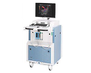
High-frequency ultrasound, designed for preclinical cardiac researchers

show detail

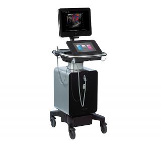
The first high-frequency, high-resolution digital imaging platform with linear array technology and Color Doppler Mode

show detail

A new and innovative platform combining ultra high-frequency ultrasound imaging, quantification and education

show detail

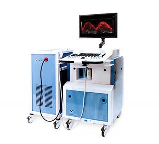
Photoacoustics is a ground-breaking, real time imaging modality for visualizing small animal anatomy and analyzing function in vivo and non-invasively

show detail

We supply and support Life-Science Technologies in the territory of Central and Eastern Europe.

Czech Republic

Slovakia

Hungary

Poland

Croatia

Slovenia

Serbia

Romania

Bulgaria

Latvia

Lithuania

Estonia

Russia

Ukraine

Belarus

Turkey