In vivo imaging

High frequency ultrasound imaging
The ultimate preclinical imaging experience!
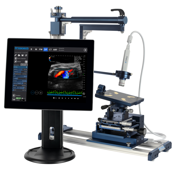
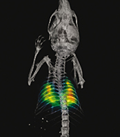
The S-Sharp high-frequency ultrasound instrument enables the researcher to obtain in vivo anatomical, functional, physiological and molecular data simultaneously, in real-time and with a resolution down to 30 µm. The system is easy to use, non-invasive and fast, providing extremely high throughput when needed.
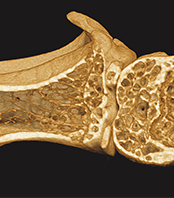
Ultrasound imaging is a well-established and validated technology that has been used clinically for many decades to visualize internal organs, soft tissues, tendons, joints and vasculature. S-Sharp has perfected the use of ultrasound in pre-clinical small animal research by creating high-frequency transducers that offers superior high resolution.
How does it work?
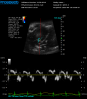
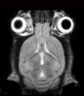
A handheld transducer is used to produce acoustic pulses that deliver sound waves into the animal’s body. Diverse tissues, organs and disease lesions absorb and reflect sound waves differently depending on their density. High-resolution grayscale images are produced when the partially reflected sound waves return to the transducer. The resulting image is viewable instantly and can be captured as a still photograph or movie.
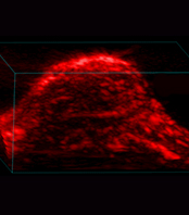
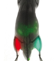
Although B-Mode, which displays a two dimensional cross-section of tissue, is the most common imaging mode with ultrasound, other image types can also be produced: blood flow, localization and direction; vascularity; tissue motion over time; presence of molecules and biomarkers; anatomy and size of a 3D region; tissue stiffness and cardiac strain.
High frequency ultrasound imaging Products
BioFocus newsletter
Register to receive Brand new technology development, Important product updates, Interesting scientific events and more!
Technologies