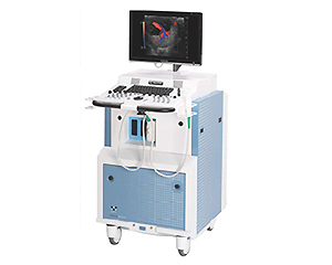
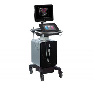
FujiFilm VisualSonics Inc. is a manufacturer of real-time, in vivo, high-resolution micro-imaging systems designed specifically for preclinical research.
Accelerate to discover
Related topics
Cardiomyocyte isolation using Cellenion’s cellenONE instrument
Mar 24, 2023
The cellenONE platform is ideal for the isolation of fragile and heterogeneous cell size populations, such as...
ACCELA educational webinar: Unlocking Spatial Biology with RareCyte Orion
Mar 16, 2023
Experience Spatial Biology with Orion Multiplex Imaging. Orion is a benchtop, high...
Recorded webinar: Anesthesia Considerations in Small Animal Imaging
Mar 13, 2023
Anesthesia settings and operation of Optical Imaging Systems: methods, animal handling, safety and regulatory...
Introducing Cytek Human Leukocyte Kit, the first ever 15-color lyse no-wash assay
Mar 13, 2023
This kit has been designed to enumerate all major leukocyte subsets and it mirrors and expands on those identified in a...
Immunofluorescence Imaging for Rare Cell Detection with CyteFinder II
Mar 7, 2023
CyteFinder II Instruments are high speed, whole slide imaging systems with options for liquid biopsy analysis and...
NanoCellect Webinar: Optimizing the Cell Line Development Process with Microfluidic Cell Sorting
Mar 3, 2023
Single-cell selection and cloning are required for bioengineering workflows such as antibody production, cell therapy,...
New Kit for Bacterial analysis expands capability of Guava Muse flow cytometer
Feb 20, 2023
With the Guava Muse cell analyzer, you can now achieve highly quantitative results at a fraction of the price, effort,...
Flash news - did you know, that you can use Singulator S100 also for plant research?
Feb 10, 2023
In this short TechNote you can find how Singulator can help the data quality in Single cell sequencing - now even in...
Jul 27, 2017
This recent article by Platt, et al. uses pulsed-wave Doppler measurement of pulmonary flow (PF) as an alternative method of cardiac output (CO) evaluation in a mouse model of myocardial infarction (MI).
Article Summary:
- Left Ventricular echocardiography remains to be a frequently used method for cardio output evaluation,
due to its non-invasive and cost efficient nature.
- Although CO derived from M-mode and B-mode images works well for healthy mice, its accuracy
decreases in certain diseased models, such as MI, where LV deformities occur.
- As an alternative, pulsed-wave Doppler flow at the pulmonary trunk was evaluated.
- CO measurements were taken from 3 echocardiography methods (M-mode, B-mode and PF) and
compared with gold standard flow probe measurements, in two diseased models (TAC and MI).
- All 3 methods correlated well with flow probe values in healthy mice, but only PF derived CO showed
strong correlation in mice with MI.
- PF measurements also showed strong intra- and inter-user agreement.
This study demonstrates that Doppler measurement of pulmonary flow may be a more robust method of CO evaluation than standard M- and B-mode measurements, especially when working with MI models. The utility of this method can be further evaluated in other cardiac disease models where LV deformation present.
Get more info
Brand profile
FujiFilm VisualSonics Inc. is a manufacturer of real-time, in vivo, high-resolution micro-imaging systems designed specifically for preclinical research.
More info at:
www.visualsonics.com