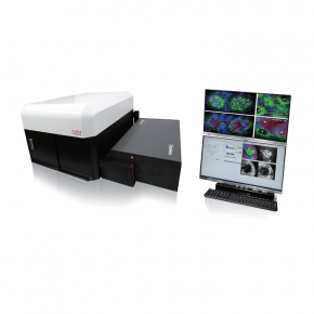
IntraVital Microscopy is a technique that enables you to directly observe the movement of live cells that make up living tissue in vivo.
Accelerate to discover
Related topics
Webinar: Multimodal tissue imaging and machine learning to advance precision medicine
May 17, 2023
Join us for this webinar to learn how the Orion spatial biology imaging platform was used to identify prognostic...
May 5, 2023
Decision to improve the output in a lab is always taken based on knowledge and workflow needs. But final decision to...
Real-Time and Quantitative Analysis of Macrophage Phagocytosis with RTCA eSight
May 4, 2023
The eSight is currently the only instrument that interrogates cell health and behavior using cellular
impedance...
Revolutionize your Flow Cytometry and Sorting workflow with Cytek Biosciences
May 3, 2023
Using full spectrum flow cytometry, Cytek systems ( RUO and CE-IVD) detect the entire fluorochrome emission, allowing...
A deep learning and Monte Carlo based framework for bioluminescence imaging center Maastro
Apr 20, 2023
"In this paper,we developed a framework using deep learning for bioluminescence-based targeting for GBM animal...
Gentle sorting of microbial cells and sub-micron particles using WOLF sorter
Apr 18, 2023
While most modern applications of flow cytometry may focus on cells of eukaryotic origin, the first flow analyzers were...
SPECT imaging properties of the CdTe-Timepix3 detector used in a new prototype
Apr 5, 2023
SPECT (Single Photon Emission Computed Tomography imaging) is one of the most important and widely used methods for the...
May 22, 2023
Chronic kidney disease (CKD) is one of the most common renal diseases manifested
by gradual loss of kidney function with no symptoms in the early stage. The underlying
mechanism in the pathogenesis of CKD with various causes such as high blood pressure, diabetes,
high cholesterol, and kidney infection is not well understood. In vivo longitudinal repetitive
cellular-level observation of the kidney of the CKD animal model can provide novel insights
to diagnose and treat the CKD by visualizing the dynamically changing pathophysiology of
CKD with its progression over time. In this study, using two-photon intravital microscopy with
a single 920 nm fixed-wavelength fs-pulsed laser, we longitudinally and repetitively observed
the kidney of an adenine diet-induced CKD mouse model for 30 days.
Continue riding on:
Article was published on 1st April 2023 in Biomedical Optics Express, Vol. 14, No. 4.

Related technologies: IntraVital Microscopy
Get more info
Brand profile
IntraVital Microscopy is a technique that enables you to directly observe the movement of live cells that make up living tissue in vivo.
More info at:
www.ivimtech.com/