Accelerate to discover

Back to filter

Related topics

Vizgen webinar: spatial relationships in Developmental Biology

Vizgen

Nov 21, 2023

MERFISH integrates spatial transcriptomics technology with high resolution spatial imaging, In this webinar we will...

ACCELA educational webinar: Drive key insights and discoveries with new live cell analysis applications for eSight

Agilent technologies

Nov 14, 2023

The xCELLigence RTCA eSight enables simultaneous real time biosensor impedance-based and...

NanoCellect webinar: Plant Potential - Gentle Cell Sorting for Enhanced Plant Biology Workflows

NanoCellect

Nov 3, 2023

Gentle cell sorting is a useful tool to increase the efficiency of plant biology workflows that include gene...

Single Cell Deposition: the cornerstone of Flow Cytometry for cellular analysis and manipulation

NanoCellect

Nov 2, 2023

One notable technology for harnessing the power of single cell deposition is the NanoCellect WOLF G2 Cell Sorter and N1...

Nov 28, 2023

Emulate in vivo conditions – introduce shear flow to your experiments with BioFlux system

Fluxion Biosciences

Oct 31, 2023

Most research is still conducted in vitro without the presence of flow. We use the BioFlux System to give you the...

Single Cell Deposition: the cornerstone of Flow Cytometry for cellular analysis and manipulation

NanoCellect

Oct 30, 2023

One notable technology for harnessing the power of single cell deposition is the NanoCellect WOLF G2 Cell Sorter and N1...

Validation of an orthotopic non-small cell lung cancer mouse model to use in radiotherapy studies

Precision X-Ray

Oct 30, 2023

The use of pre-clinical orthotopic mouse models is essential for the development of novel therapies targeting solid...

Nov 28, 2023

Atypical Teratoid Rhabdoid Tumours Are Susceptible to Panobinostat-Mediated Differentiation Therapy

Spectral Instruments Imaging

Oct 20, 2023

To assess tumour growth, mice were imaged using the AMI-HTX bioluminescent imaging system (Spectral Instruments...

Show all topics (10)

High-plex immunofluorescence imaging and traditional histology of the same tissue section

Aug 7, 2023

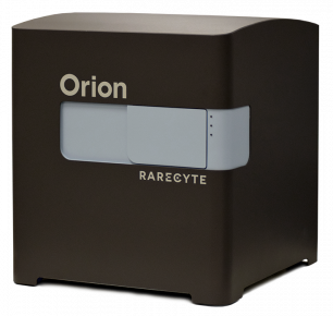
Precision medicine is critically dependent on better methods for diagnosing and staging disease and predicting drug response. Histopathology using hematoxylin and eosin (H&E)-stained tissue (not genomics) remains the primary diagnostic method in cancer. Recently developed highly multiplexed tissue imaging methods promise to enhance research studies and clinical practice with precise, spatially resolved single-cell data. Here, we describe the ‘Orion’ platform for collecting H&E and high-plex immunofluorescence images from the same cells in a whole-slide format suitable for diagnosis. Using a retrospective cohort of 74 colorectal cancer resections, we show that immunofluorescence and H&E images provide human experts and machine learning algorithms with complementary information that can be used to generate interpretable, multiplexed image-based models predictive of progression-free survival. Combining models of immune infiltration and tumor-intrinsic features achieves a 10- to 20-fold discrimination between rapid and slow (or no) progression, demonstrating the ability of multimodal tissue imaging to generate high-performance biomarkers.

Learn more
Scientific paper
Application

Get more info

Riccardo Pasculli

Head of application support

Riccardo

Pasculli

+420 731 127 717

Send Message

Brand profile

RareCyte

The whole-slide, highly multiplexed biomarker imaging platform - depth and flexibility with the convenience of a comprehensive, rapid, single cycle process.

Related products

The benchtop, high resolution whole-slide fluorescence imaging system that enables rapid spatial insights

show detail

We supply and support Life-Science Technologies in the territory of Central and Eastern Europe.

Czech Republic

Slovakia

Hungary

Poland

Croatia

Slovenia

Serbia

Romania

Bulgaria

Latvia

Lithuania

Estonia

Russia

Ukraine

Belarus

Turkey